ARG55558
anti-ATP5B antibody
anti-ATP5B antibody for Flow cytometry,ICC/IF,IHC-Formalin-fixed paraffin-embedded sections,Western blot and Human
Controls and Markers antibody; Metabolism antibody; Signaling Transduction antibody
概述
| 产品描述 | Rabbit Polyclonal antibody recognizes ATP5B |
|---|---|
| 反应物种 | Hu |
| 预测物种 | Ms, Rat, Bov |
| 应用 | FACS, ICC/IF, IHC-P, WB |
| 宿主 | Rabbit |
| 克隆 | Polyclonal |
| 同位型 | IgG |
| 靶点名称 | ATP5B |
| 抗原物种 | Human |
| 抗原 | KLH-conjugated synthetic peptide corresponding to aa. 135-163 (Center) of Human ATP5B. |
| 偶联标记 | Un-conjugated |
| 別名 | ATPMB; ATPSB; EC 3.6.3.14; HEL-S-271; ATP synthase subunit beta, mitochondrial |
应用说明
| 应用建议 |
|
||||||||||
|---|---|---|---|---|---|---|---|---|---|---|---|
| 应用说明 | * The dilutions indicate recommended starting dilutions and the optimal dilutions or concentrations should be determined by the scientist. | ||||||||||
| 阳性对照 | WiDr |
属性
| 形式 | Liquid |
|---|---|
| 纯化 | Purification with Protein A and immunogen peptide. |
| 缓冲液 | PBS and 0.09% (W/V) Sodium azide |
| 抗菌剂 | 0.09% (W/V) Sodium azide |
| 存放说明 | For continuous use, store undiluted antibody at 2-8°C for up to a week. For long-term storage, aliquot and store at -20°C or below. Storage in frost free freezers is not recommended. Avoid repeated freeze/thaw cycles. Suggest spin the vial prior to opening. The antibody solution should be gently mixed before use. |
| 注意事项 | For laboratory research only, not for drug, diagnostic or other use. |
生物信息
| 数据库连接 |
Swiss-port # P06576 Human ATP synthase subunit beta, mitochondrial |
|---|---|
| 基因名称 | ATP5B |
| 全名 | ATP synthase, H+ transporting, mitochondrial F1 complex, beta polypeptide |
| 背景介绍 | This gene encodes a subunit of mitochondrial ATP synthase. Mitochondrial ATP synthase catalyzes ATP synthesis, utilizing an electrochemical gradient of protons across the inner membrane during oxidative phosphorylation. ATP synthase is composed of two linked multi-subunit complexes: the soluble catalytic core, F1, and the membrane-spanning component, Fo, comprising the proton channel. The catalytic portion of mitochondrial ATP synthase consists of 5 different subunits (alpha, beta, gamma, delta, and epsilon) assembled with a stoichiometry of 3 alpha, 3 beta, and a single representative of the other 3. The proton channel consists of three main subunits (a, b, c). This gene encodes the beta subunit of the catalytic core. [provided by RefSeq, Jul 2008] |
| 生物功能 | Mitochondrial membrane ATP synthase (F(1)F(0) ATP synthase or Complex V) produces ATP from ADP in the presence of a proton gradient across the membrane which is generated by electron transport complexes of the respiratory chain. F-type ATPases consist of two structural domains, F(1) - containing the extramembraneous catalytic core, and F(0) - containing the membrane proton channel, linked together by a central stalk and a peripheral stalk. During catalysis, ATP synthesis in the catalytic domain of F(1) is coupled via a rotary mechanism of the central stalk subunits to proton translocation. Subunits alpha and beta form the catalytic core in F(1). Rotation of the central stalk against the surrounding alpha(3)beta(3) subunits leads to hydrolysis of ATP in three separate catalytic sites on the beta subunits. [UniProt] |
| 细胞定位 | Mitochondrion. Mitochondrion inner membrane. Note=Peripheral membrane protein |
| 研究领域 | Controls and Markers antibody; Metabolism antibody; Signaling Transduction antibody |
| 预测分子量 | 57 kDa |
检测图片 (4) Click the Picture to Zoom In
-
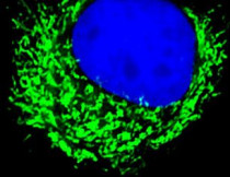
ARG55558 anti-ATP5B antibody ICC/IF image
Immunofluorescence: SK-BR-3 cells stained with ARG55558 anti-ATP5B antibody (green) at 1:25 dilution. DAPI (blue) for nuclear staining.
-
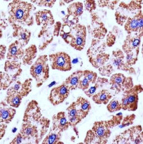
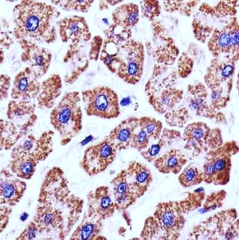
ARG55558 anti-ATP5B antibody IHC-P image
Immunohistochemistry: Paraffin-embedded Human liver section stained with ARG55558 anti-ATP5B antibody at 1:25 dilution.
-

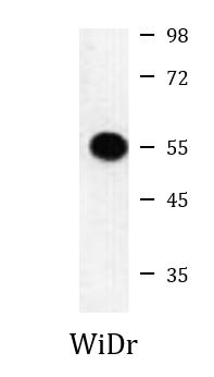
ARG55558 anti-ATP5B antibody WB image
Western blot: WiDr cell lysate stained with ARG55558 anti-ATP5B antibody.
-
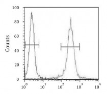
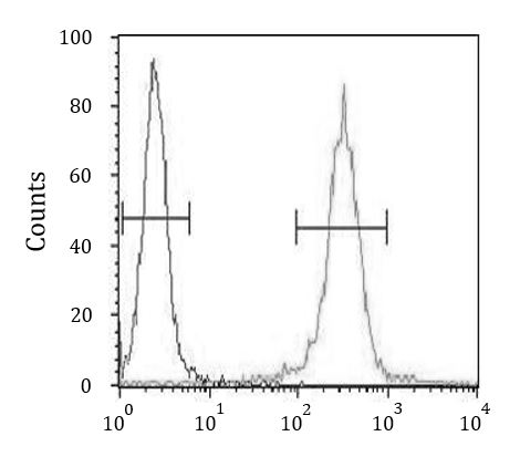
ARG55558 anti-ATP5B antibody FACS image
Flow Cytometry: WiDr cells stained with ARG55558 anti-ATP5B antibody (right histogram) or without primary antibody control (left histogram), followed by incubation with FITC labelled secondary antibody.