ARG30106
Mitochondria / Caspase Dependant Apoptosis Marker Antibody Duo (Caspase9, Cytochrome c)
Cancer antibody; Cell Biology and Cellular Response antibody; Cell Death antibody; Metabolism antibody; Signaling Transduction antibody
内含物
| 货号 | 内含物名称 | 宿主克隆性 | 反应 | 应用 | 包装 |
|---|---|---|---|---|---|
| ARG20048 | anti-Caspase 9 (cleaved) antibody | Rabbit pAb | Hu | IP, WB | 25 μg |
| ARG20006 | anti-Cytochrome C antibody | Rabbit pAb | Hu, Ms, Rat, Bov, Chk, Dog | IP, WB | 25 μg |
概述
| 产品描述 | Cytochrome C is an electron transport protein localized in the mitochondria. When a cell receive intra- or extra-cellular death signal, Cytochrome C is then released from mitochondria to cytoplasm and trigger Procaspase 9 activation. Detection of the cytoplasmic release of Cytochrome C and activation of Caspase 9 with anti-Cytochrome C (ARG20006) and anti-Caspase 9 (ARG51317) antibodies are early indications that a cell is undergoing apoptosis. |
|---|---|
| 靶点名称 | Mitochondria / Caspase Dependant Apoptosis Marker |
| 別名 | Mitochondria/Caspase Dependant Apoptosis Marker antibody; Cytochrome C antibody; Caspase 9 (cleaved) antibody |
属性
| 存放说明 | For continuous use, store undiluted antibody at 2-8°C for up to a week. For long-term storage, aliquot and store at -20°C or below. Storage in frost free freezers is not recommended. Avoid repeated freeze/thaw cycles. Suggest spin the vial prior to opening. The antibody solution should be gently mixed before use. |
|---|---|
| 注意事项 | For laboratory research only, not for drug, diagnostic or other use. |
生物信息
| 全名 | Antibody Duo for Mitochondria/Caspase Dependant Apoptosis Marker (Caspase9, Cytochrome c) |
|---|---|
| 产品亮点 | Related Product: anti-Cytochrome C antibody; anti-Dopamine Transporter antibody; |
| 研究领域 | Cancer antibody; Cell Biology and Cellular Response antibody; Cell Death antibody; Metabolism antibody; Signaling Transduction antibody |
检测图片 (4) Click the Picture to Zoom In
-
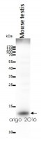
ARG20006 anti-Cytochrome C antibody WB image
Western blot: 20 µg of Mouse testis lysate stained with ARG20006 anti-Cytochrome C antibody at 2 µg/ml dilution.
-
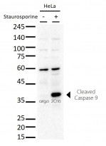
ARG20048 anti-Caspase 9 (cleaved) antibody WB image
Western blot: 20 µg of HeLa untreated or treated with Staurosporine and stained with ARG20048 anti-Caspase 9 (cleaved) antibody at 1:500 dilution.
-
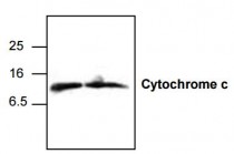
ARG20006 anti-Cytochrome c antibody WB image
Western Blot: 1. Jurkat cell lysate 2. 3T3 cell lysate stained with anti-Cytochrome c antibody (ARG20006).
The cytochrome c antibody detects the 12.6 kDa cytochrome c from human, mouse, and rat samples. Jurkat cell lysate, NIH3T3 cell lysate, and rat kidney tissue lysate can be used as positive controls. -
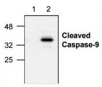
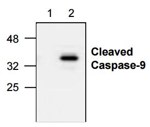
ARG20048 anti-Caspase-9 (Active) antibody WB image
Western Blot: 1. Untreated Jurkat cell lysate 2. Etoposide treated Jurkat cell lysate stained with anti-Caspase 9 active antibody (ARG20048).
The anti-active caspase-9 antibody recognizes only the cleaved caspase-9 (37 kDa). It does not recognize full-le ngth caspase-9 or any other caspases.


文献引用
Single-Cell Analysis of Signaling Proteins Provides Insights into Proapoptotic Properties of Anticancer Drugs.
ARG20006: scPISA / Human


Probing apoptosis signaling proteins in single living cells for precision efficacy evaluation of anti-cancer drugs.
ARG20006: / Human


